The Dry Eye Center at
How we diagnose dry eye disease.
We Can Help Dry Eye!
Dry eye syndrome affects millions of Americans, presenting symptoms that vary from occasional, mild irritation to persistent and challenging issues. It's crucial to recognize that dry eye disease is often a chronic and progressive condition, demanding well-thought-out maintenance protocols and continuous tracking. In this rapidly evolving field, our commitment is to stay at the forefront by integrating the newest treatment technologies and recently approved pharmacologic treatments. Our goal is to enhance the quality of life and visual functioning for our patients dealing with dry eye.
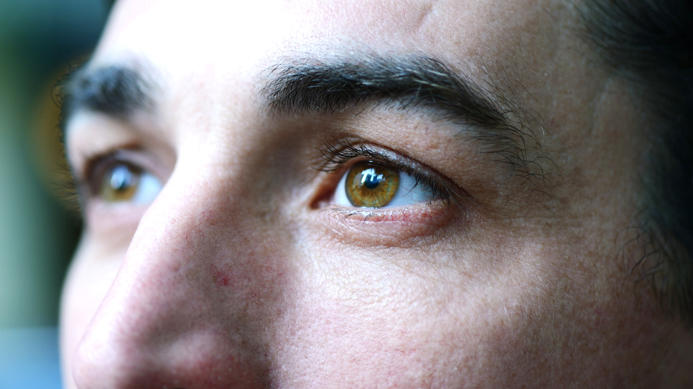
Each blink spreads tears across the cornea, providing essential lubrication, reducing infection risk, and washing away foreign matter. Unusual as it may seem, an increase in tears or an appearance of crying can signal dry eyes. If you suspect dry eye symptoms or simply want a thorough examination of your glands, schedule an appointment with our dedicated dry eye specialist, Dr. Alan Margolis.
Dr. Alan Margolis conducts a Comprehensive History, delving into current medications, previous medical conditions, including autoimmune diseases, smoking history, and sleep apnea. With a proprietary scoring system, he integrates various metrics to tailor specific treatment protocols for each patient. Embrace the dynamic landscape of dry eye management with our expertise, ensuring optimal care and continuous adaptation to the latest advancements.
Dr Margolis has treated over 45,000 patients in the past 34 years.
Dry Eye Diagnostics
How Does Dr. Margolis Diagnosis Dry Eye?
1. SPEED questionnaire
Diagnosising Ocular Surface Disease (dry eye) begins with an initial questionairre. The SPEED questionnaire was designed in order to quickly track the progression of dry eye symptoms over time. This questionnaire gives a score from 0 to 28 that is the result of 8 items that assess frequency and severity of symptoms.

2. Corneal Topography
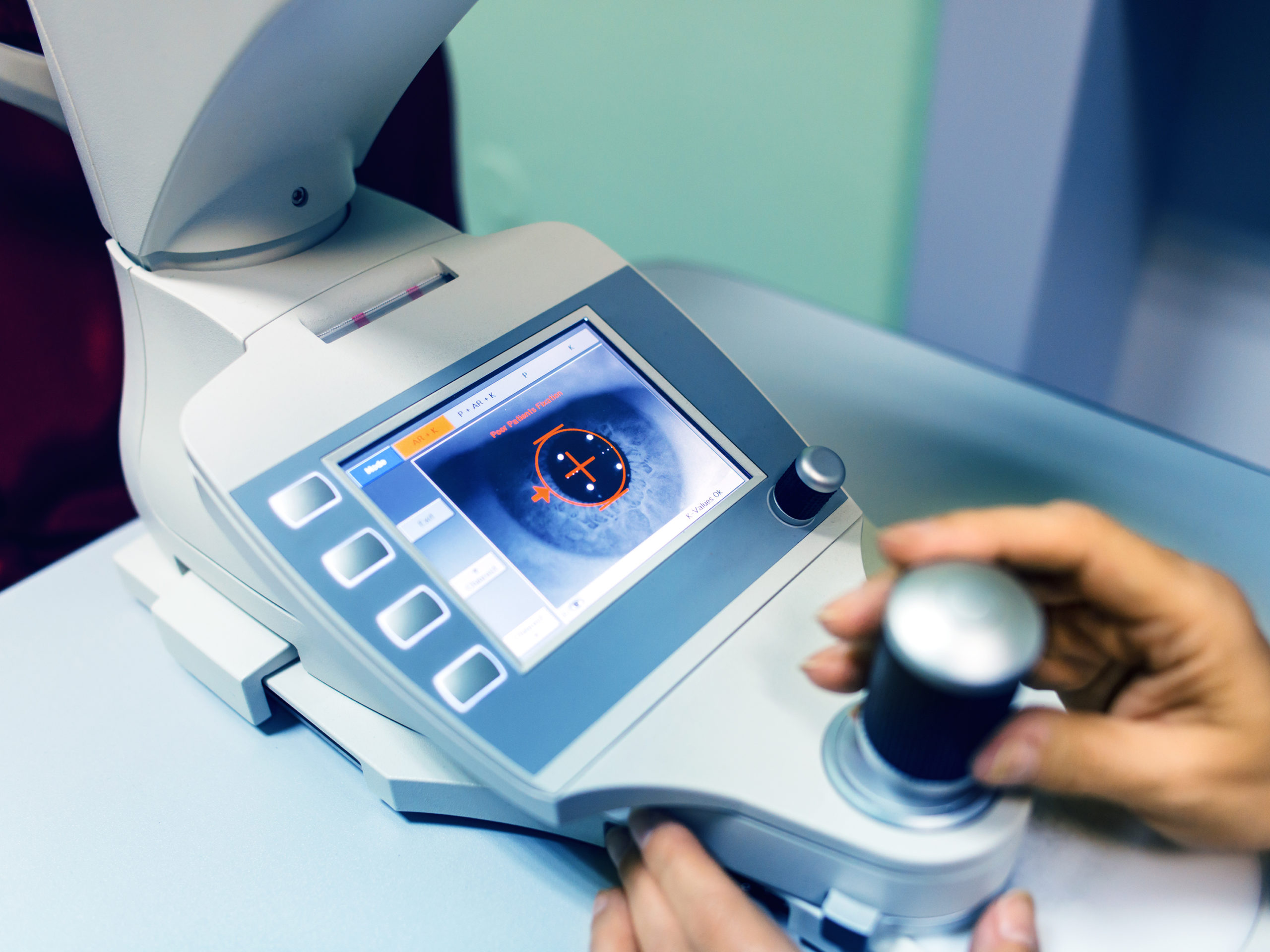
Corneal topography is a special photography technique that maps the surface of the clear, front window of the eye (the cornea). It works much like a 3D (three-dimensional) map of the world, that helps identify features like mountains and valleys. But with a topography scan, a doctor can find distortions in the curvature of the cornea, which is normally smooth. It also helps doctors monitor eye disease and plan for surgery.
Getting a corneal topography is painless, as nothing touches your eye during the scan.
3. Lipiscan
LipiScan, a non-invasive procedure, enables Dr Margolis to view the health of your meibomian glands with optimum accuracy. This diagnostic tool has changed the way medicine diagnoses dry eye syndrome. With efficiency and versatility, LipiScan is a small and lightweight device with a comfortable patient experience. Quick and painless, the procedure takes about one minute to image both lower eyelids.
More info here
More info here
More info here
More info here
More info here
4. Lipiview
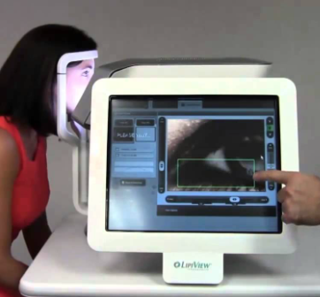
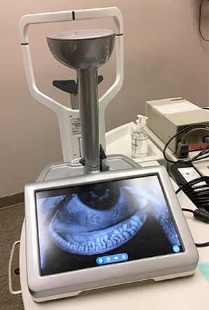
LipiView is an innovative, digital imaging tool used to evaluate the severity of a patient’s dry eye disease and meibomian gland dysfunction (MGD). Dr Margolis will use this evaluation to measure your eyes’ natural lipid production in your tear layer, blink rate, incomplete blinking, and determine if you have Meibomian Gland Dysfunction (MGD), a blockage of your eyes’ oil producing glands. Your doctor is then able to customize your treatment regimen based on the severity.
LipiView is done in office and only takes about 5 minutes. You will simply look into a camera and blink normally. You will be able to see pictures of your own glands, and Dr Margolis will be able to determine whether you have Meibomian Gland Dysfunction and dry eye disease. LipiView can also evaluate your blinking function by assessing if your blinks are complete. Incomplete blinking results in a poor-quality tear film which can contribute to ongoing dry eye symptoms.
Include Lipid layer thickness
